


1
2
3
4
5
6
7
8
9
10
11
12
13
14
15
16
Blood flow through a normal aorta and an aorta with an aortic dissection
The blood flow in a healthy aorta compared to the blood flow in an aortic dissection. In a dissection, a tear in the inner layer of the vessel causes blood to flow between the layers and form a false lumen. Depicted here is a type A aortic dissection - a dissection occurring in the ascending aorta.
Ankle sprain
A sprained ankle occurs when you roll or twist your ankle, causing the ligaments around your ankle to become damaged. This illustration depicts some of the ligaments that can be torn in a severe ankle sprain, including the anterior tibiofibular ligament anterior talofibular ligament, and calcaneofibular ligament.
Cross sectional anatomy of the eye
Cross section anatomy of the eye, including the optic nerve, fovea, sclera, choroid, retina, lens, zonular fibers, pupil, cornea, pars plicata, and pars plana.
Three component model of force transmission in muscles
Diagram showing the three component model of force transmission in muscles, including the parallel elastic component (PEC), series elastic component (SEC), and contractile component (CE). The contractile element is further made up of sarcomeres containing the microfilaments actin and myosin, while titin is an element of the series elastic component.
Compression in long and short muscle lengths
Compression in Long and Short Muscle Lengths
Diagram comparing the compression of the three component of force transmission in a biceps brachii muscle in long and short muscle length positions.
DNA structure
Diagram of the structure of DNA, including the major groove, minor groove, sugar phosphate backbone, and base pairs of adenine, thymine, guanine, and cytosine.
Sagittal view of the cervical spine
Illustration showing the anatomy of a sagittal section of the cervical spine.
Anatomy of the kidneys
Illustration depicting the anatomy a pair of kidneys, including the ureter, and a cross-section showing the renal pelvis, renal cortex, renal medulla, major calyx, and minor calyx.
Lobes of the brain
Illustration detailing the major regions of the brain, including the frontal lobe, parietal lobe, temporal lobe, occipital lobe, and cerebellum.
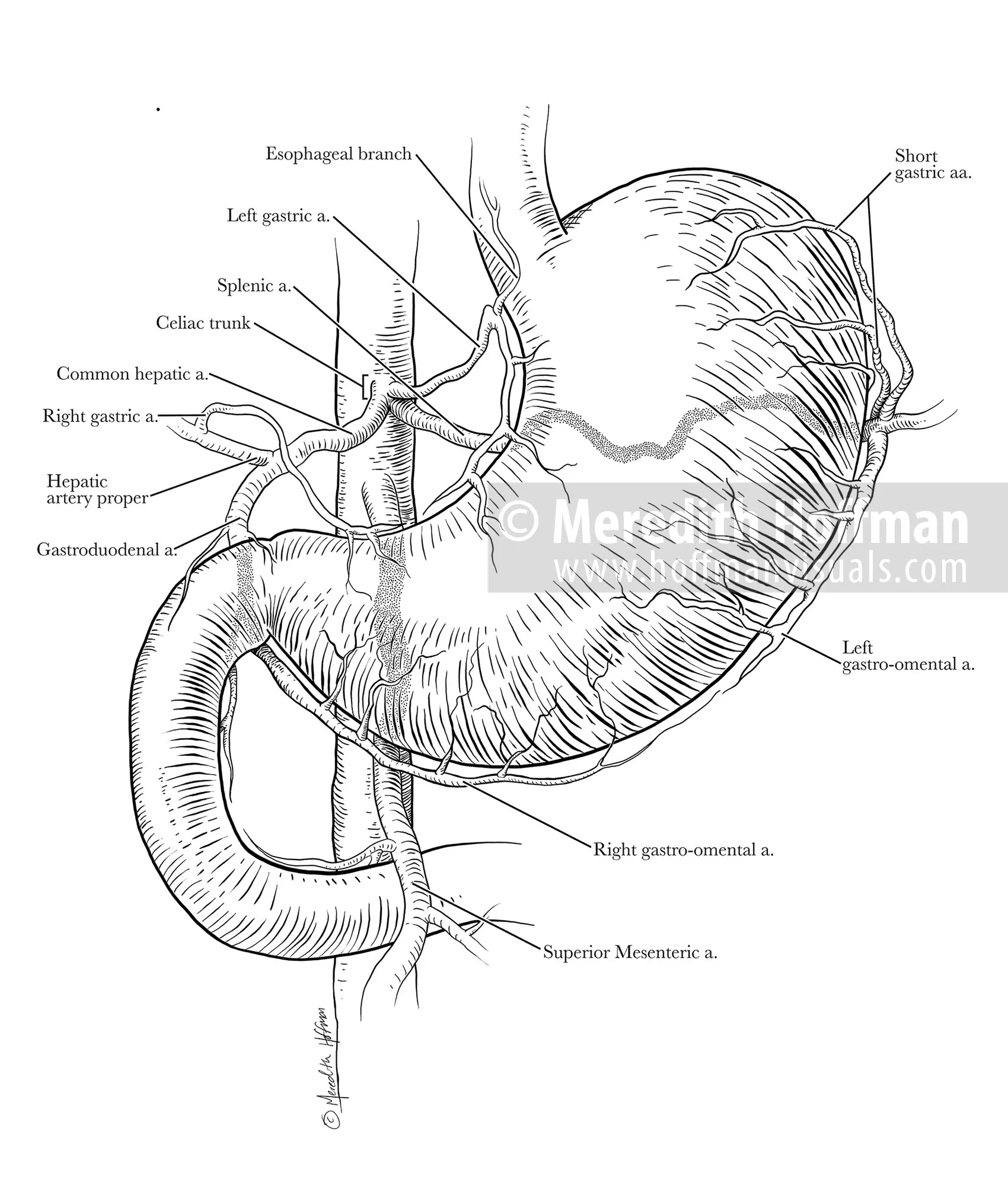
Celiac trunk and blood supply to the stomach
Anatomy of the blood supply to the stomach and duodenum, originating from the celiac trunk and abdominal aorta. Branches include the left gastric artery, esophageal branch, splenic artery, short gastric arteries, left gastro-omental artery, common hepatic artery, hepatic artery proper, right gastric artery, gastroduodenal artery, right gastro-omental artery, and the superior mesenteric artery.
Anatomy of the shoulder and its ligaments
Illustration depicting the anterior anatomy of the glenohumeral joint, including the humerus, clavicle, scapula, coracoacromial ligament, acromioclavicular ligament, coracoclavicular ligament, trapezoid ligament, and conoid ligament.
Eczema of the foot
Editorial illustration depicting eczema of the foot. This illustration originally accompanied a writer’s description of life with this condition, published in Esopus Magazine.
Typhoid at the Chicago 1893 World's Fair
Published as a cover illustration for the Northwestern Public Health Review, accompanying Dr. Bronwyn Rae's article on the typhoid outbreak in Chicago leading up to the Columbian Exposition
Surgical instruments
Line illustrations showing the structure of several common surgical instruments, including a retractor, hemostat, and a gloved hand with scissors.
Anatomy of the heart
Illustration showing the anatomy of the anterior heart, including the aorta, vena cava, pulmonary vessels, and coronary vessels.
Blood flow through the heart
Cross section of the heart with arrows showing the path that blood flows through the vena cava, right atrium, right ventricle, pulmonary artery, left atrium, left ventricle, and aorta.